
What is it?
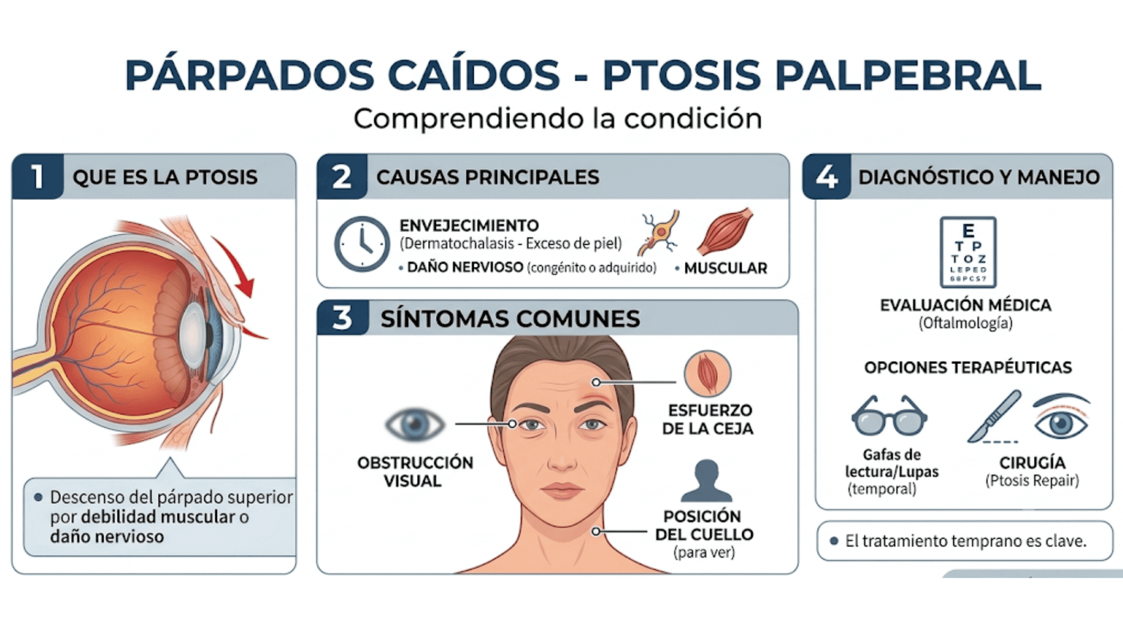
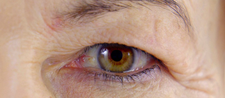
Palpebral ptosis, or a drooping eyelid, is an abnormal downward displacement of the upper eyelid—covering the eye partially or completely—caused by weakness of the levator muscle. It can be congenital (present at birth) or acquired with age, and it may affect vision and the visual field, as well as cause aesthetic concerns.
Among the various types of ptosis, the most common in children is simple congenital palpebral ptosis. Another, much rarer form of congenital ptosis is known as blepharophimosis syndrome. Marcus-Gunn syndrome and Horner syndrome are other forms of palpebral ptosis. Mechanical or traumatic ptosis may also occur.
Causes
The most common cause is aging of the levator muscle (aponeurotic). However, it can also be congenital, result from a lack of nerve stimulation (known as neurogenic), or be caused by the presence of cysts and/or tumors (known as mechanical).
Symptoms
Eyelid ptosis can be mild (the drooping eyelid partially covers the upper part of the pupil), moderate, or severe, and symptoms may include:
- Malposition of the eyelid that affects the visual axis.
- Reduced vision.
- Eye fatigue.
- Elevation of the eyebrows.
- The need to tilt the head back to see better.
How do we treat it?
Ptosis (drooping eyelid) is primarily corrected through outpatient surgery performed under local anesthesia, designed to strengthen or shorten the eyelid’s levator muscle. Surgical approaches include anterior (external) techniques, transconjunctival (scarless) techniques, or frontalis suspension; the specific method is chosen based on the severity of the drooping and the function of the muscle.
A specialized oculoplastic evaluation is essential to determine the underlying cause and the most appropriate surgical technique, as impaired muscle function may require a different type of procedure. Main Surgical Procedures
- Strengthening/Shortening of the Levator Muscle: This is the most common technique when muscle function is good. The tension of the levator muscle is adjusted to lift the eyelid.
- Anterior Approach: A small incision is made within the natural crease of the eyelid to access the muscle, which allows the resulting scar to remain hidden.
- Transconjunctival Approach (Putterman technique): The muscle is strengthened from the inner side of the eyelid, leaving no external scar.
- Frontal Suspension: Used in severe cases where the levator muscle is barely functional (severe or congenital ptosis). The eyelid is connected to the frontalis muscle (in the eyebrow area), allowing the patient to open their eyes by raising their eyebrows.
- Whitnall’s Ligament Sling Procedure: Specifically designed for severe ptosis, this technique uses Whitnall’s ligament as an upper support structure.