
Inicio - SERVICES - Ophthalmology - Cornea and ocular surface: Anterior segment - Corneal transplant
What is it?
A corneal transplant is a surgery in which a patient’s diseased cornea is replaced with a healthy cornea from a donor. The cornea is the clear front part of the eye that covers the colored iris and the round pupil. Light is focused as it passes through the cornea, allowing us to see. To remain clear, the cornea must be healthy.
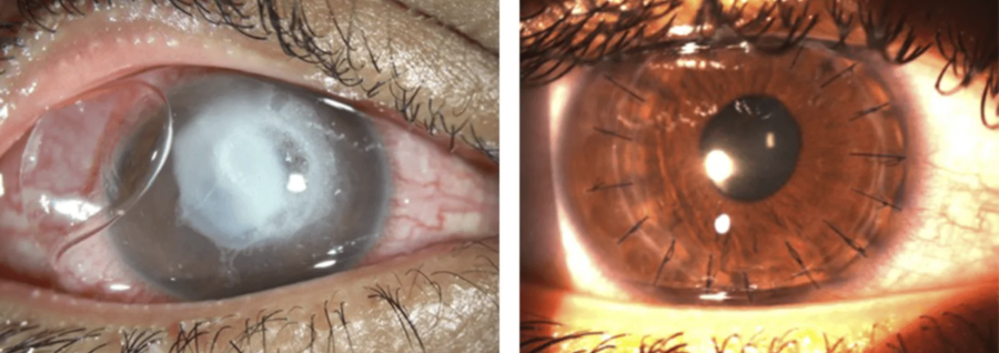
If the cornea is damaged, it may become swollen or scarred. In either case, it may lose its smooth, clear appearance. Scarring, swelling, or an irregular shape of the cornea causes light to scatter or become distorted, resulting in blurred vision or the appearance of halos.
A corneal transplant is necessary if:
Rejection following an initial corneal transplant.
Scarring following an injury.
- There is corneal damage following another eye surgery, such as cataract surgery.
- There is keratoconus, a pronounced curvature of the cornea.
- There is a hereditary corneal disease, such as Fuchs’ dystrophy.
How do we treat it?
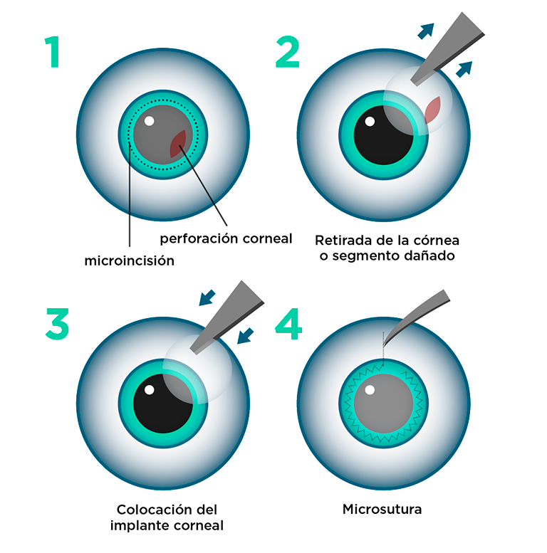
A corneal transplant is a procedure that lasts approximately 45 to 90 minutes, depending on the complexity of the case. To undergo the procedure, the patient must be registered with an organ bank while awaiting a donor. The procedure is painless and is performed under local or general anesthesia, depending on the patient’s age, medical condition, and the specific eye disease.
Depending on which part of the tissue is affected, the specialist may perform a full-thickness transplant (penetrating keratoplasty)—in which the entire damaged cornea is replaced with tissue from a healthy donor—or a lamellar transplant —in which only the affected layers (such as the endothelium or the anterior layers) are replaced, allowing for faster visual recovery. Once the diseased or damaged cornea has been carefully removed from the eye, the new, clear cornea is sutured into place.
Although the rejection rate for corneal transplants in cases of keratoconus ranges from 5% to 30%—with rejection leading to impaired or blurred vision—most instances of rejection can be halted with minimal damage, provided they are treated in a timely manner. For inflammatory eye diseases, the rejection rate is higher.
Warning signs of rejection include persistent discomfort, sensitivity to light, red eyes, or changes in vision; other potential complications include infection, bleeding, swelling, retinal detachment, or glaucoma. Any of these symptoms should be reported to the treating physician, as all such complications can be addressed. A corneal transplant can be performed again—often with good results—though the overall risk of rejection is higher for a second transplant than for the first.
In some cases, the SLET (Simple Limbal Epithelial Transplantation) technique is used to reconstruct the ocular surface in patients with limbal stem cell deficiency—a condition in which the cells responsible for regenerating the cornea and keeping it transparent are damaged or destroyed.