

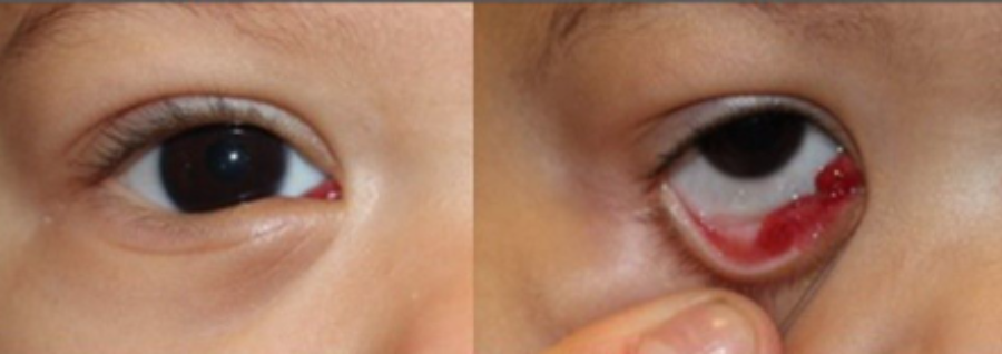
What are they?
These are benign (non-cancerous) or malignant (cancerous) masses that develop within the eyeball or the surrounding tissues. While many are congenital and benign, malignant neoplasms can pose a serious risk to a child’s vision and life.
Pediatric ocular tumors are classified as follows:
- Retinoblastoma: an aggressive cancer that begins in the retina; it can affect one or both eyes.
- Capillary hemangioma: a benign tumor (the most common type in infants) resulting from the proliferation of blood vessels.
- Dermoid cyst and lymphangioma: cystic lesions or benign vascular malformations that often appear as periorbital masses.
Symptoms
Sometimes these are difficult to notice at first glance, but the most common warning signs include:
- Leukocoria: A white reflection in the pupil, often noticeable when taking a photo with a flash (instead of the typical red-eye effect).
- Strabismus: Eyes that are misaligned or do not look in the same direction.
- Changes in the eye: Persistent redness, swelling, pain, or changes in the color of the iris.
- Vision loss: The child may frequently trip or fail to follow objects with their gaze.
Causes
Most of these tumors occur due to genetic mutations—errors in the DNA of eye cells—that cause the cells to multiply uncontrollably. In some cases, such as with certain retinoblastomas, these mutations can be inherited from one’s parents; however, in the vast majority of instances, they occur entirely sporadically and randomly during a child’s development, and cannot be prevented.
How do we treat them?
The primary goal is to save the child’s life, followed by preserving as much vision as possible. The main medical treatments include:
- Chemotherapy: Special medications used to shrink or eliminate tumor cells. It can be administered systemically or injected directly into the arteries leading to the eye (intra-arterial chemotherapy) to minimize side effects in the rest of the body.
- Focal therapies: For small tumors, extreme cold (cryotherapy) or heat from a laser (thermotherapy) is used to destroy the affected tissue.
- Enucleation
In this procedure, the surgeon carefully removes the affected eye containing the tumor, ensuring that a portion of the optic nerve is also removed to prevent any diseased cells from remaining. During the same operation, an internal sphere (implant) is placed into the eye socket, and the eye muscles are attached to it to preserve as much movement as possible.
The surgery is performed under general anesthesia and typically lasts between 1 and 2 hours.
Following the procedure, the patient will need to wear a pressure bandage over the eye for a few days. Additionally, antibiotic and anti-inflammatory drops or ointments must be applied, and the patient should be kept on relative bed rest—avoiding rough play, contact sports, or any activities that could result in a blow to the area—for several weeks.Between 4 and 6 weeks after surgery, once the area has completely healed, the child is referred to a prosthetics specialist to design a custom-made artificial eye that is aesthetically identical to the healthy eye.