

What is it?
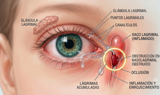
Lacrimal passage stenosis (or a blocked tear duct) is the narrowing or obstruction of the channel that connects the eye to the nose. Normally, tears cleanse the eye and drain through this internal “conduit.” When the duct is blocked, tears have nowhere to go and instead accumulate, causing them to constantly overflow.
Symptoms
The most common signs that can be observed on a daily basis are:
- Constant tearing (epiphora): The eye appears excessively moist, or tears may be seen running down the cheek, even when the child is not crying.
- Frequent eye discharge: A thick, yellowish or greenish discharge accumulates—especially upon waking—because the stagnant fluid easily becomes infected.
- Irritated skin: The area around the eyelids or the cheek may become red due to constant moisture.
- Mild swelling: A small lump near the inner corner of the eye (the lacrimal sac).
Causes
In children, the primary cause is congenital. This means they are born with an incompletely developed tear duct. Most commonly, a small, thin membrane at the end of the duct (known as the valve of Hasner) has failed to open properly before birth.
How do we treat her?
In nearly 90% of cases, following the attending physician’s recommendations, the condition resolves on its own during the first year of life. The ophthalmologist may initially recommend a specific technique—known as the Crigler massage—to help push fluid through and break the small, obstructing membrane. It is also essential to maintain strict hygiene by cleaning away eye discharge using sterile gauze and saline solution. In cases where there are clear signs of infection (such as significant green or yellow discharge), the doctor may also prescribe antibiotic eye drops.
If the problem persists after the child turns one (or sooner, if infections are particularly severe), a pediatric ophthalmologist may suggest a simple procedure known as nasolacrimal duct probing.
- Nasolacrimal Duct Probing: This procedure is performed under short-acting general inhalation anesthesia to ensure the child remains still, comfortable, and free from pain or anxiety. It typically lasts between 10 and 15 minutes. During the procedure, the specialist guides a probe through the tear duct—reaching the back of the nasal cavity—to break the thin membrane causing the obstruction. They then perform tests to confirm that the duct is completely open.
This is an outpatient procedure, and the child will be prescribed antibiotic and anti-inflammatory eye drops to be used for one to two weeks. It is normal for the child to experience mild nosebleeds or pink-tinged tears during the first few days; however, they can generally resume their normal activities (such as eating and playing) almost immediately.